
Cerebellum
Page most recently updated 7 September 2026
Lesions of the cerebellum can produce both ataxia and psychiatric manifestations. Ataxia ➚ is explored in more detail elsewhere on this site, from the perspective of the movement lens ➚. This page focuses on the psychiatric, cognitive, and behavioural manifestations associated with cerebellar lesions, collectively referred to as cerebellar cognitive affective syndrome (CCAS). CCAS encompasses executive, language, visuospatial, and affective disturbances and is particularly associated with lesions affecting posterior cerebellar regions.
Psychiatric, cognitive, and behavioural effects of lesions may also arise through mechanisms other than network disruption; the potential contribution of these mechanisms can be considered using the Neuropsychiatric Effects of CNS Structural Abnormalities (NECSA) classification.
On this page:
Anterior lobe
Flocculonodular lobe
Posterior lobe
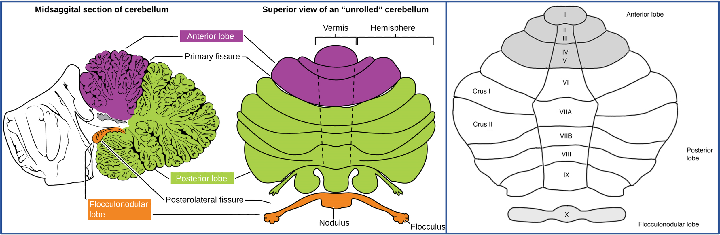
Cerebellar lobes and lobules (per Larsell’s classification)
The anterior lobe consists of lobules I–V, the posterior lobe consists of lobules VI–IX (with Crus I and Crus II constituting the most lateral hemispheric parts of lobule VIIa), and the flocculonodular lobe consists of lobule X.
Image credit: Left image adapted from OpenStax College via Wikimedia Commons. Licensed under the Creative Commons Attribution 3.0 International (CC BY 3.0): https://creativecommons.org/licenses/by/3.0/. Right image adapted from Malte S. Depping, Mike M. Schmitgen, Katharina M. Kubera and Robert C. Wolf via Wikimedia Commons. Licensed under the Creative Commons Attribution 4.0 International (CC BY 4.0): https://creativecommons.org/licenses/by/4.0/. Composite figure created by combining images from the above sources. Title and caption modified.
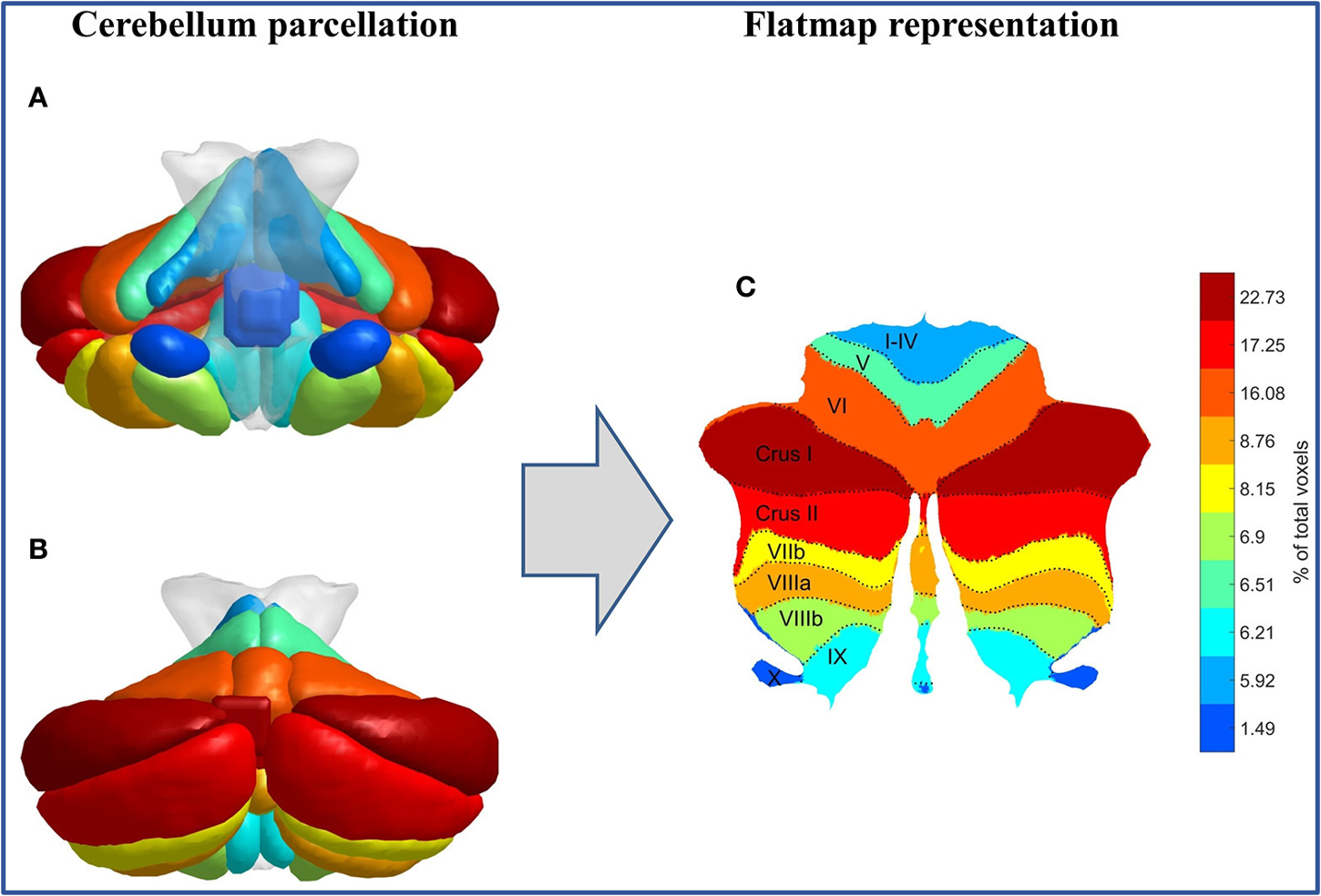
Cerebellar lobes and lobules in 3D
A: Coronal view, anterior; B: Coronal view, posterior; C: Flat surface representation.
Image credit: Adapted from Vasileios C. Pezoulas, Michalis Zervakis, Sifis Michelogiannis and Manousos A. Klados via Wikimedia Commons. Licensed under the Creative Commons Attribution 4.0 International (CC BY 4.0): https://creativecommons.org/licenses/by/4.0/. Title and caption modified.
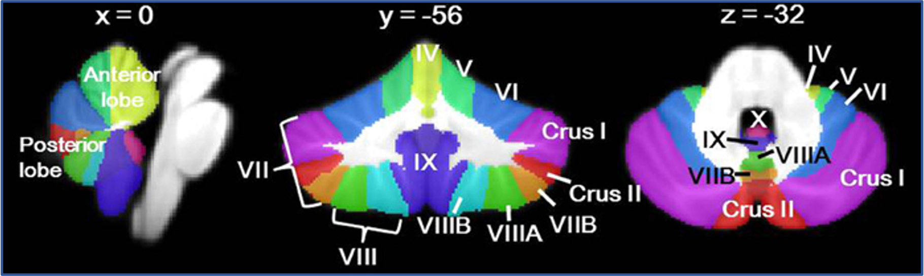
Cerebellar lobes and lobules on MRI
Sagittal, coronal and axial planes, from left to right.
Image credit: Adapted from Catherine J. Stoodley via Wikimedia Commons. Licensed under the Creative Commons Attribution 3.0 International (CC BY 3.0): https://creativecommons.org/licenses/by/3.0/. Title and caption modified.
1. Anterior lobe → Cerebellar motor syndrome
Hemispheres
• Ipsilateral limb ataxia
Vermis and paravermal regions
• Truncal ataxia
2. Flocculonodular lobe → Vestibulocerebellar syndrome
• Oculomotor deficits, including nystagmus
• Impaired balance
3. Posterior lobe → Cerebellar cognitive affective syndrome (CCAS; Schmahmann syndrome)
CCAS is characterized by four major groups of symptoms:
1. Executive dysfunction
• Impaired planning, set-shifting and abstraction
• Associated primarily with lesions in lobules VII and VIII
2. Language impairment
• Agrammatism and impaired fluency
• Dysprosodia
• Associated primarily with lesions in right posterolateral cerebellar regions (right Crus I, Crus II and lobule IX)
3. Affective and behavioural changes
• Personality changes
• Blunting of affect and social withdrawal
• Disinhibited behaviour and irritability
• Possibly more strongly associated with lesions in vermal regions
4. Visuospatial impairment
• Simultanagnosia-like features
• Visuospatial disorganization and impaired visuospatial memory
• Associated primarily with lesions in bilateral Crus I, Crus II and right lobule VIII
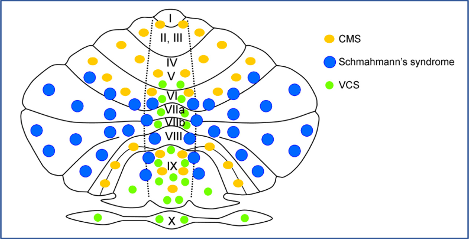

Localization of cerebellar syndromes across lobules
The illustration shows an unfolded cerebellum, with the three cerebellar syndromes localized to lobules labelled according to Larsell's classification. The syndromes are cerebellar motor syndrome (CMS), cerebellar cognitive affective syndrome (CCAS; also known as Schmahmann's syndrome), and vestibulocerebellar syndrome (VCS).
Image credit: Adapted from Manto M, Mariën P. Schmahmann's syndrome – identification of the third cornerstone of clinical ataxiology. Cerebellum & Ataxias. 2015;2:2. https://doi.org/10.1186/s40673-015-0023-1. Licensed under the Creative Commons Attribution 4.0 International (CC BY 4.0): https://creativecommons.org/licenses/by/4.0/. Title and caption modified.
Lesion maps of cerebellar syndromes in sagittal, coronal and axial planes
Individual lesion maps from patients presenting with (A) cerebellar motor syndrome only, (B) cerebellar cognitive affective syndrome (CCAS) only, and (C) both cerebellar motor syndrome and CCAS. Different colours represent lesions from individual patients. The deep cerebellar nuclei (fastigial, interposed and dentate) are shown in violet, red and yellow, respectively. This image can be compared with the previous figure, “Cerebellar lobes and lobules on MRI,” which provides a reference map of cerebellar regions.
Image credit: Stoodley C et al. Location of lesion determines motor vs. cognitive consequences in patients with cerebellar stroke. NeuroImage: Clinical. 2016;12:765–775. https://doi.org/10.1016/j.nicl.2016.10.013. Licensed under the Creative Commons Attribution-NonCommercial-NoDerivatives 4.0 International (CC BY-NC-ND 4.0): https://creativecommons.org/licenses/by-nc-nd/4.0/. Image used unchanged. Website title and accompanying description modified.
Clinical reminder: consider NECSA mechanisms
(Neuropsychiatric Effects of CNS Structural Abnormalities)
When a structural lesion is identified in a patient presenting with neuropsychiatric symptoms, consider which NECSA mechanisms may explain these symptoms:
● Network disruption
Disruption of functional networks due to the presence of the lesion
● Electrical disturbance
Ictal, preictal, postictal, or interictal effects due to epileptic activity associated with the lesion (particularly if involving or adjacent to cortex)
● Chemical (endocrine) effects
Hormone excess or deficiency caused by the lesion (particularly if involving the pituitary gland or hypothalamus)
● Side effects of treatment
Effects of medications, surgery, radiotherapy, or other interventions
● Alternative explanations
The lesion may be incidental, or symptoms may arise from another cause (e.g., psychological response to illness or a comorbid psychiatric condition)
Selected references and further reading — Lesional lens
Rohlfing T, Zahr NM, Sullivan EV, Pfefferbaum A. The SRI24 multichannel atlas of normal adult human brain structure. Hum Brain Mapp. 2010;31(5):798–819.
Clark DL, Boutros NN, Mendez MF. The brain and behavior: an introduction to behavioral neuroanatomy. 4th ed. Cambridge: Cambridge University Press; 2018.
Brazis PW, Masdeu JC, Biller J. Localization in clinical neurology. 7th ed. Philadelphia: Wolters Kluwer/Lippincott Williams & Wilkins; 2016.
Blumenfeld H. Neuroanatomy through clinical cases. 3rd ed. New York: Oxford University Press; 2021.
Hassan I. The neuropsychiatric effects of CNS structural abnormalities (NECSA) classification: an aid to differential diagnosis. Aust N Z J Psychiatry. 2015;49(10):943.
Arciniegas DB, Yudofsky SC, Hales RE, editors. The American Psychiatric Association Publishing textbook of neuropsychiatry and clinical neurosciences. 6th ed. Washington (DC): American Psychiatric Association Publishing; 2018.
Agrawal N, Faruqui R, Bodani M, editors. Oxford textbook of neuropsychiatry. Oxford: Oxford University Press; 2020.