
Fusiform gyrus
Page most recently updated 7 September 2026
This page focuses on the neuropsychiatric manifestations associated with lesions of the fusiform gyrus.
Cognitive, behavioural, and psychiatric effects of lesions may also arise through mechanisms other than network disruption; the potential contribution of these mechanisms can be considered using the Neuropsychiatric Effects of CNS Structural Abnormalities (NECSA) classification.
On this page:
● Identification using the standardized 15-slice MRI atlas
● Lesion manifestations
● Clinical pearls
● Identification using the standardized 15-slice MRI atlas
The fusiform gyrus is identified by a translucent colour overlay in the relevant slices of the standardized 15-slice MRI atlas. For an explanation of the slices specifically curated for the atlas across the three anatomical planes, together with unmarked MRI images, please see the atlas’s dedicated page ➚.
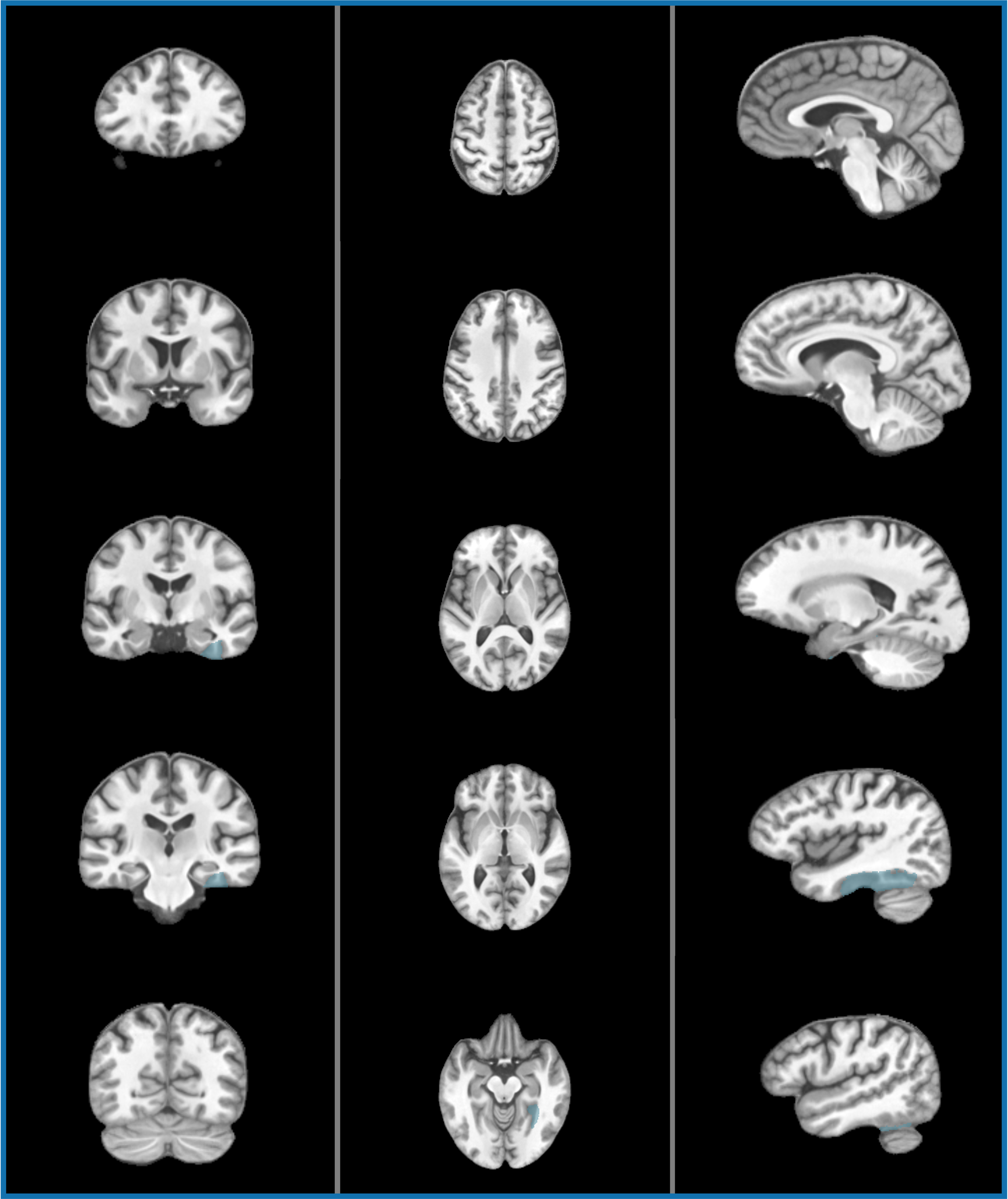
Fusiform gyrus - MRI atlas
The fusiform gyrus is identified by a translucent colour overlay in the relevant T1-weighted slices of the standardized 15-slice MRI atlas, across the coronal, axial and sagittal planes.
Image credit: MRI adapted from the SRI24 Atlas (Rohlfing et al., 2010), CC BY-SA 3.0; anatomical overlays generated using the atlas’s SRI24/LPBA40 label map.
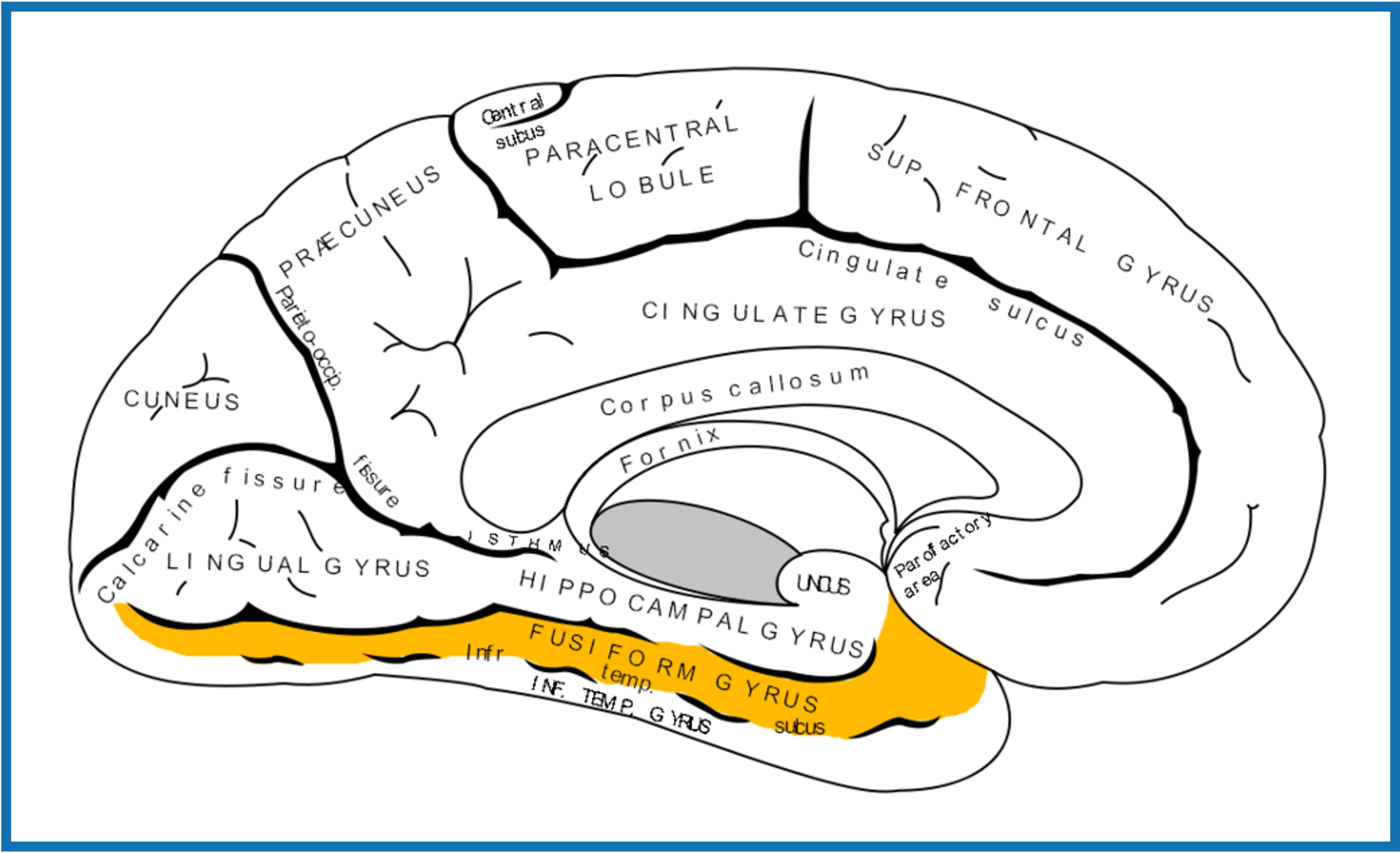
Fusiform gyrus
Diagram of the cerebrum, with the fusiform gyrus marked in yellow.
Image credit: Adapted from Gray's Anatomy, via Wikimedia Commons. Public domain.
● Lesion manifestations
▪ Right fusiform involvement:
◦ Prosopagnosia
◦ Impaired recognition of familiar faces
◦ Reduced facial familiarity
▪ Left fusiform involvement:
◦ Alexia without agraphia (with splenial involvement)
◦ Impaired visual word recognition
▪ Bilateral involvement:
◦ Visual agnosia
● Clinical pearls
▪ Fusiform lesions are among the most clinically recognisable examples of selective cognitive deficits following focal brain injury.
▪ Patients with prosopagnosia often retain:
◦ Voice recognition
◦ Emotional familiarity
◦ Knowledge that a face is a face
▪ The deficit is usually not an inability to see or recognise a face as a face, but an impaired ability to link the visual percept with a specific identity.
Clinical reminder: consider NECSA mechanisms
(Neuropsychiatric Effects of CNS Structural Abnormalities)
When a structural lesion is identified in a patient presenting with neuropsychiatric symptoms, consider which NECSA mechanisms may explain these symptoms:
● Network disruption
Disruption of functional networks due to the presence of the lesion
● Electrical disturbance
Ictal, preictal, postictal, or interictal effects due to epileptic activity associated with the lesion (particularly if involving or adjacent to cortex)
● Chemical (endocrine) effects
Hormone excess or deficiency caused by the lesion (particularly if involving the pituitary gland or hypothalamus)
● Side effects of treatment
Effects of medications, surgery, radiotherapy, or other interventions
● Alternative explanations
The lesion may be incidental, or symptoms may arise from another cause (e.g., psychological response to illness or a comorbid psychiatric condition)
Selected references and further reading — Lesional lens
Rohlfing T, Zahr NM, Sullivan EV, Pfefferbaum A. The SRI24 multichannel atlas of normal adult human brain structure. Hum Brain Mapp. 2010;31(5):798–819.
Clark DL, Boutros NN, Mendez MF. The brain and behavior: an introduction to behavioral neuroanatomy. 4th ed. Cambridge: Cambridge University Press; 2018.
Brazis PW, Masdeu JC, Biller J. Localization in clinical neurology. 7th ed. Philadelphia: Wolters Kluwer/Lippincott Williams & Wilkins; 2016.
Blumenfeld H. Neuroanatomy through clinical cases. 3rd ed. New York: Oxford University Press; 2021.
Hassan I. The neuropsychiatric effects of CNS structural abnormalities (NECSA) classification: an aid to differential diagnosis. Aust N Z J Psychiatry. 2015;49(10):943.
Arciniegas DB, Yudofsky SC, Hales RE, editors. The American Psychiatric Association Publishing textbook of neuropsychiatry and clinical neurosciences. 6th ed. Washington (DC): American Psychiatric Association Publishing; 2018.
Agrawal N, Faruqui R, Bodani M, editors. Oxford textbook of neuropsychiatry. Oxford: Oxford University Press; 2020.