
Pituitary region
Page most recently updated 7 September 2026
Lesions of the sellar and suprasellar region can produce psychiatric, cognitive, and behavioural manifestations through multiple mechanisms. Pituitary adenomas and craniopharyngiomas are considered separately because the relative contribution of these mechanisms differs: endocrine effects are particularly prominent in functioning pituitary adenomas, whereas network disruption from involvement of adjacent hypothalamic and basal forebrain structures is particularly characteristic of craniopharyngiomas. This distinction illustrates the importance of considering multiple pathogenic mechanisms using the Neuropsychiatric Effects of CNS Structural Abnormalities (NECSA) classification.
On this page:
1. Pituitary adenomas
2. Craniopharyngiomas
3. Other lesions of the pituitary region
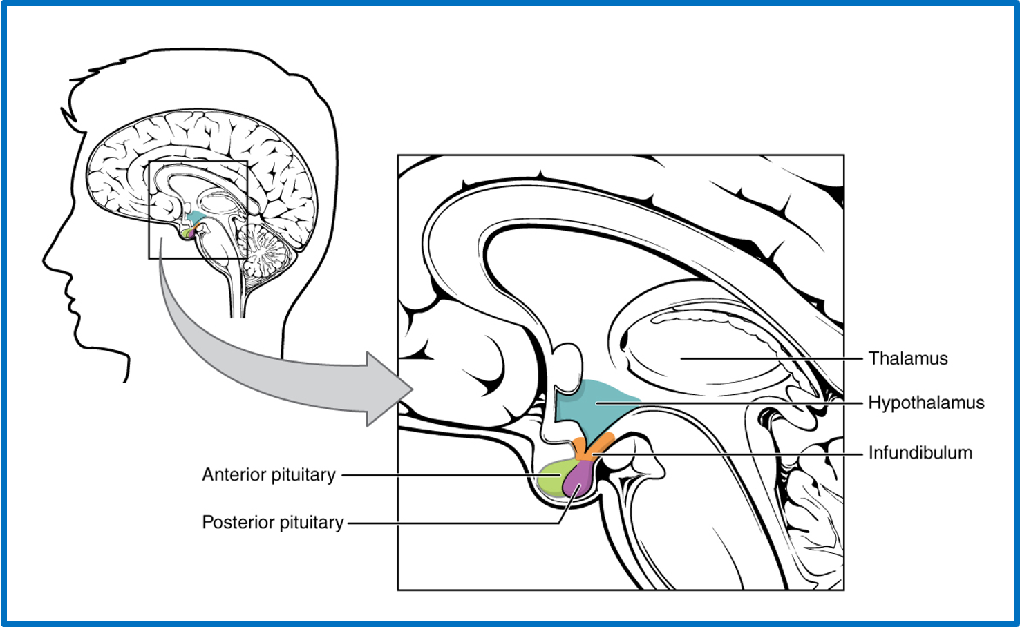
Pituitary gland and suprasellar anatomy
Illustration of the pituitary gland and suprasellar region in the sagittal plane.
Image credit: OpenStax College, The Hypothalamus–Pituitary Complex, from Anatomy & Physiology. Licensed under the Creative Commons Attribution 3.0 Unported (CC BY 3.0) licence. Title and caption modified.
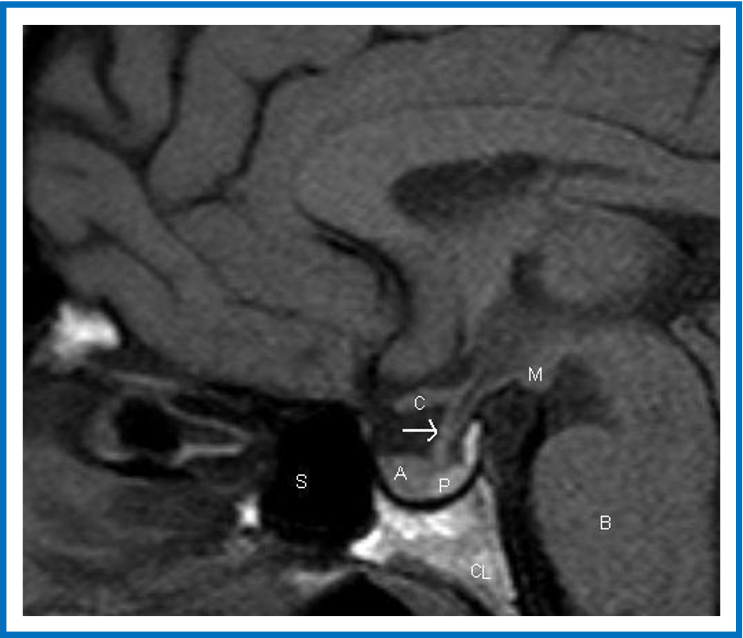
MRI of the normal pituitary gland - sagittal view
Sagittal T1-weighted unenhanced image of the pituitary fossa demonstrates normal anatomy. The anterior pituitary tissue (A) is visible within the sella, and the posterior pituitary bright spot (P) is evident behind it. The stalk (arrow) is well seen, with a small cleft of CSF visible within it superiorly — the infundibular recess of the third ventricle. The optic chiasm (C) and mammillary bodies (M) are seen in the suprasellar region. B, brainstem; S, sphenoid air sinus; CL, clivus.
Image credit: Evanson J. Radiology of the Pituitary. In: Feingold KR, Adler RA, Ahmed SF, et al., editors. Endotext [Internet]. South Dartmouth (MA): MDText.com, Inc.; 2000–. Last Update: September 14, 2023. Available from NCBI Bookshelf. Licensed under the Creative Commons Attribution-NonCommercial-NoDerivatives 2.0 (CC BY-NC-ND 2.0) licence.
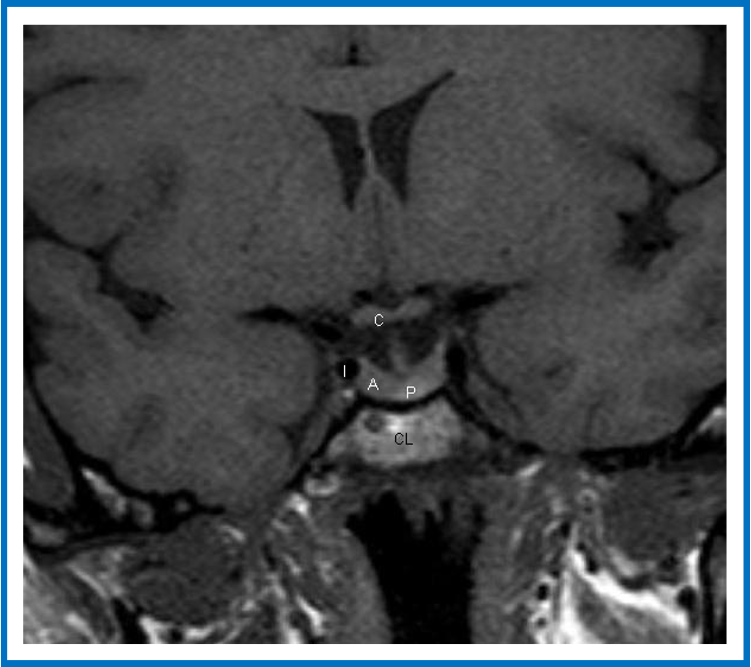
MRI of the normal pituitary gland - coronal view
Coronal T1-weighted unenhanced image of the pituitary fossa demonstrating normal anatomy. The anterior pituitary gland (A) is within the fossa. The posterior pituitary bright spot (P) is visible centrally—a normal finding on T1-weighted MRI corresponding to the posterior pituitary (neurohypophysis). The stalk is seen extending superiorly into the suprasellar region. The optic chiasm (C) is visible. The cavernous segments of the carotid arteries (I) are seen within the cavernous sinuses, which form the lateral boundaries of the pituitary fossa.
Image credit: Evanson J. Radiology of the Pituitary. In: Feingold KR, Adler RA, Ahmed SF, et al., editors. Endotext [Internet]. South Dartmouth (MA): MDText.com, Inc.; 2000–. Last Update: September 14, 2023. Available from NCBI Bookshelf. Licensed under the Creative Commons Attribution-NonCommercial-NoDerivatives 2.0 (CC BY-NC-ND 2.0) licence.
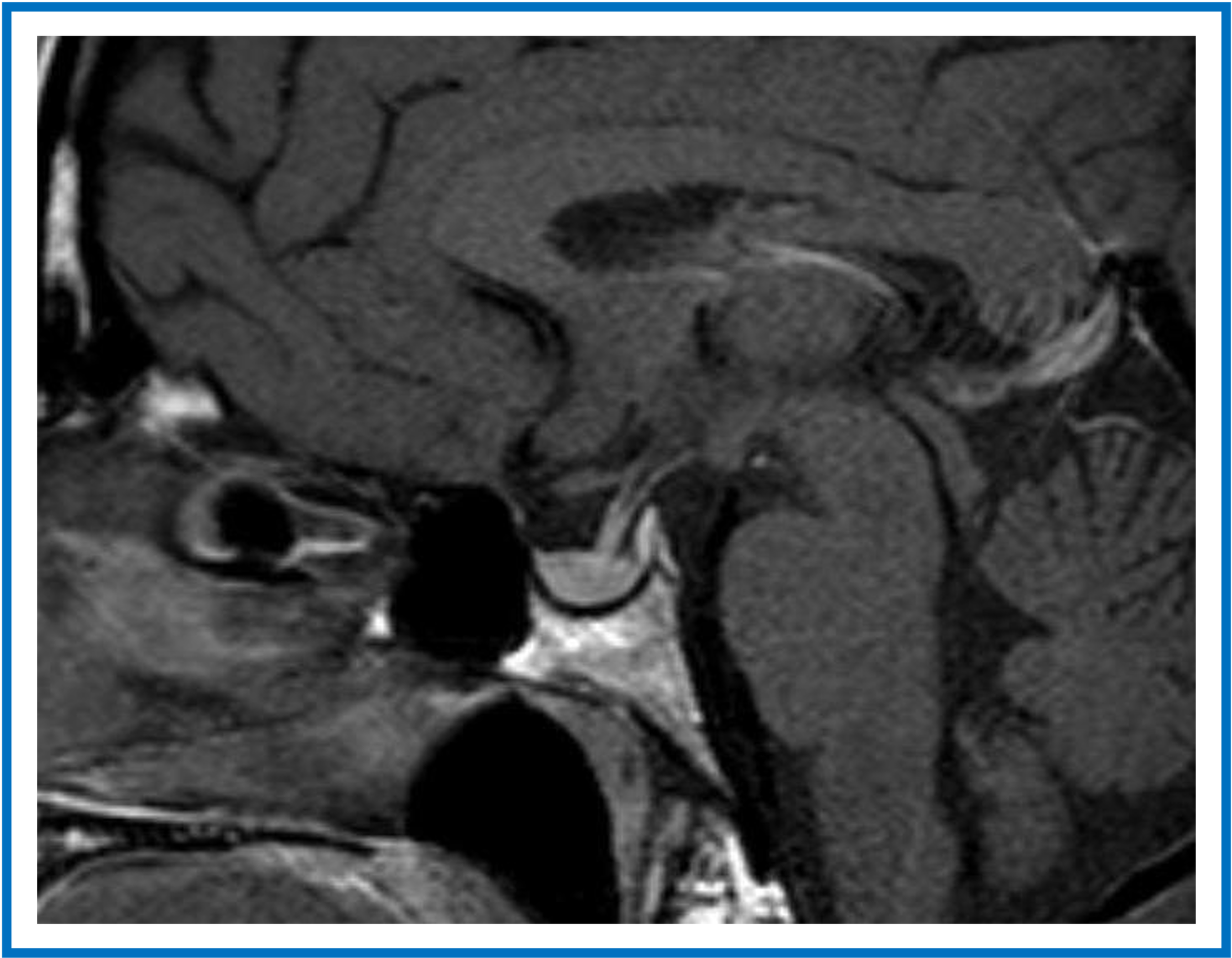
MRI of the normal pituitary gland — postcontrast sagittal view
Sagittal T1-weighted postcontrast image of the pituitary. The pituitary gland and pituitary stalk demonstrate normal contrast enhancement.
Image credit: Evanson J. Radiology of the Pituitary. In: Feingold KR, Adler RA, Ahmed SF, et al., editors. Endotext [Internet]. South Dartmouth (MA): MDText.com, Inc.; 2000–. Last Update: September 14, 2023. Available from NCBI Bookshelf. Licensed under the Creative Commons Attribution-NonCommercial-NoDerivatives 2.0 (CC BY-NC-ND 2.0) licence.
1. Pituitary adenomas
Pituitary adenomas (also known as pituitary neuroendocrine tumours, or PitNETs, in the current WHO classification) may produce psychiatric manifestations through endocrine disturbance, mass effect, or treatment-related effects.
ACTH-secreting adenomas (Cushing disease) are the pituitary tumours most strongly associated with psychiatric manifestations. Chronic hypercortisolism commonly causes depression, anxiety, emotional lability, and cognitive impairment, and less commonly mania or psychosis.
Other functioning adenomas may produce psychiatric symptoms indirectly through endocrine disturbance. Prolactinomas are among the most common functioning pituitary adenomas and may affect mood, wellbeing, and sexual function through hyperprolactinaemia and associated hypogonadism. Somatotroph adenomas may indirectly contribute to psychiatric symptoms through the systemic effects of growth hormone excess, while TSH-secreting adenomas are rare but may produce symptoms related to secondary hyperthyroidism.
Large pituitary macroadenomas may compress normal pituitary tissue, resulting in hypopituitarism. Secondary adrenal insufficiency, hypothyroidism, and hypogonadism may contribute to depression, apathy, cognitive impairment, and reduced libido.
Behavioural disturbance due to tumour mass effect is uncommon but may occur with giant suprasellar lesions extending into the hypothalamus and adjacent basal forebrain. Such patients may develop apathy, abulia, executive dysfunction, and impaired motivation through disruption of frontal-subcortical motivational networks.
Treatment of prolactinomas with dopamine agonists, particularly cabergoline, may rarely produce neuropsychiatric adverse effects, including impulse-control disorders (especially pathological gambling, compulsive shopping, binge eating, and hypersexuality), mood elevation, mania, or psychosis.
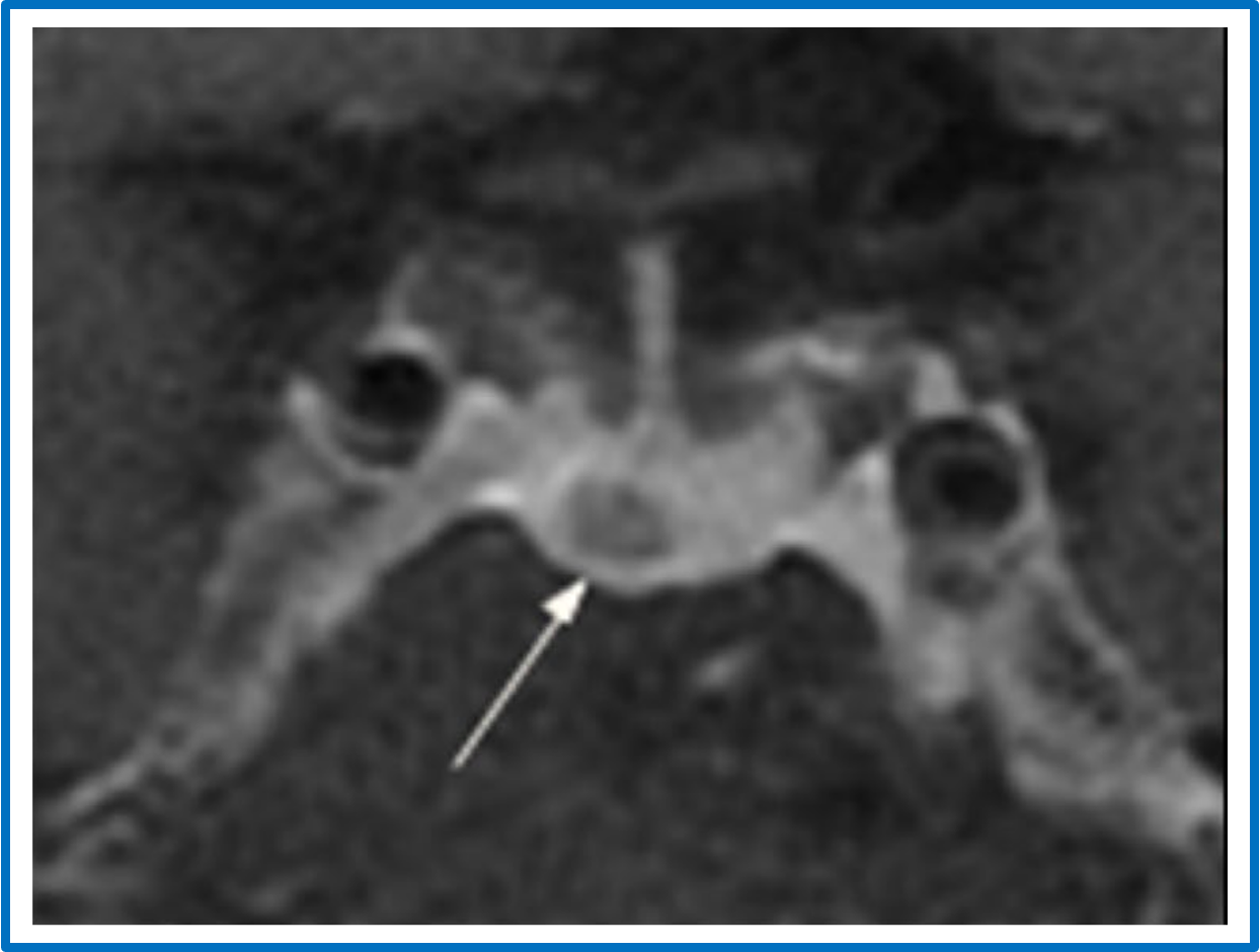
Pituitary microadenoma
Coronal contrast-enhanced T1-weighted high-resolution MRI demonstrating a 5-mm pituitary microadenoma (arrow) identified using a three-dimensional fast spin echo (3D FSE) sequence.
Image credit: Adapted from Liu Z, Hou B, You H, et al. High-resolution contrast-enhanced MRI with three-dimensional fast spin echo improved the diagnostic performance for identifying pituitary microadenomas in Cushing’s syndrome. European Radiology. 2023;33:5984–5992. doi:10.1007/s00330-023-09585-1. Licensed under the Creative Commons Attribution 4.0 International (CC BY 4.0) licence. Modifications have been made.
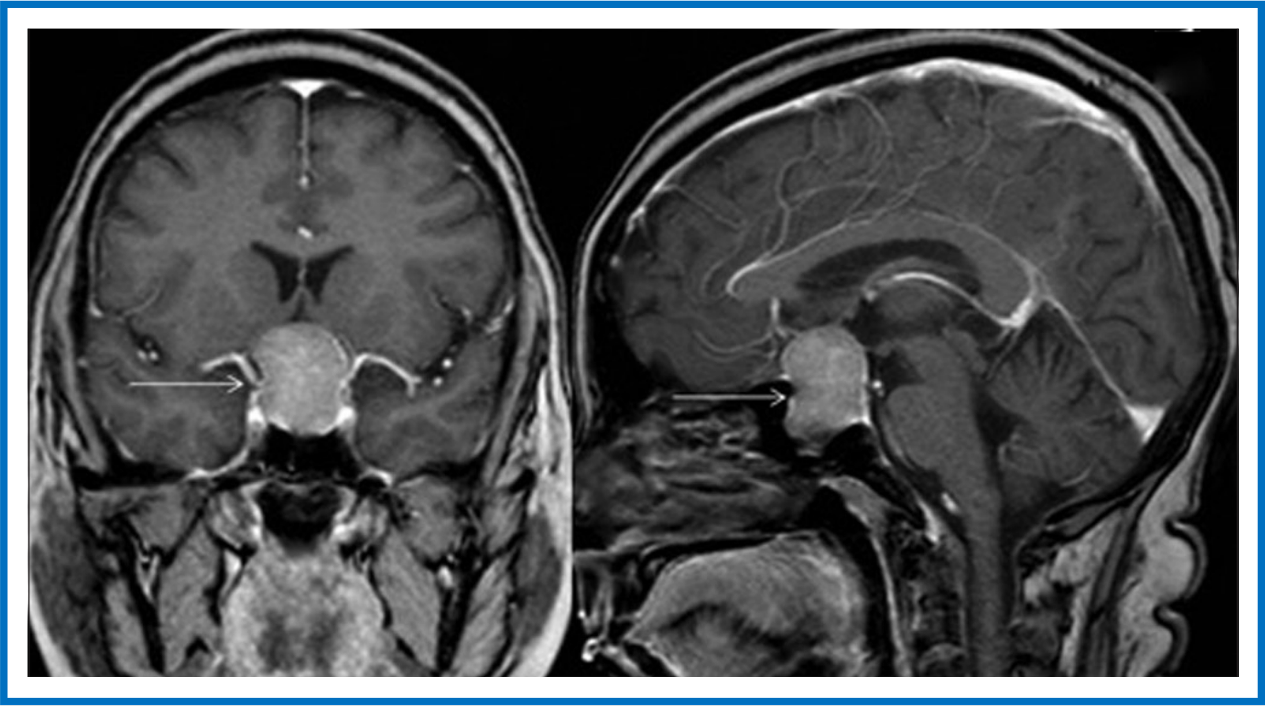
Pituitary macroadenoma
Large inhomogeneous pituitary macroadenoma (arrows) demonstrating marked enhancement on postcontrast coronal (left) and sagittal (right) images.
Image credit: Adapted from Chaudhary V, Bano S. Imaging of the pituitary: Recent advances. Indian Journal of Endocrinology and Metabolism. 2011;15(Suppl 3):S216–S223. doi:10.4103/2230-8210.84871. Licensed under the Creative Commons Attribution-NonCommercial-ShareAlike 3.0 Unported (CC BY-NC-SA 3.0) licence. Modifications include cropping the original six-panel figure to retain the coronal and sagittal postcontrast MRI images (original panels C and D) and removal of panel labels. Title and caption modified.
2. Craniopharyngiomas
Craniopharyngiomas are rare tumours of the sellar and suprasellar region but are disproportionately important in neuropsychiatry because they arise adjacent to the hypothalamus, pituitary stalk, and basal forebrain.
Compared with pituitary adenomas, craniopharyngiomas are considerably more likely to produce prominent behavioural and cognitive disturbances through direct involvement of hypothalamic–limbic networks, in addition to endocrine dysfunction.
Adult presentations commonly include executive dysfunction, apathy, cognitive impairment, hypersomnolence, hypopituitarism, and visual impairment. In children, presentation more commonly includes growth failure, delayed puberty, hypothalamic obesity, headache, and visual impairment, with persistent neurocognitive and behavioural sequelae frequently developing following treatment, reflecting both the effects of the tumour and treatment-related injury to hypothalamic networks.
Hypothalamic involvement may result in hypothalamic syndrome, characterized by disturbances of appetite, sleep–wake regulation, autonomic and endocrine function, motivation, cognition, and behaviour (see Hypothalamus).
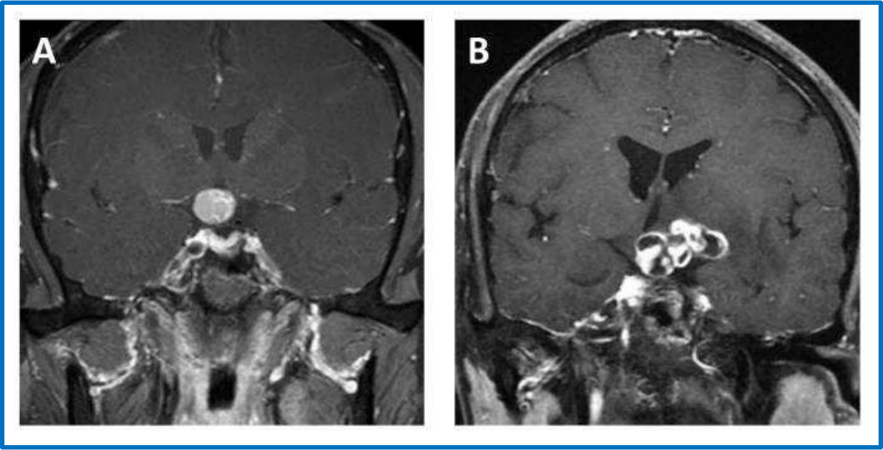
Craniopharyngiomas
Contrast-enhanced T1-weighted coronal MR images of (A) adamantinomatous craniopharyngioma and (B) papillary craniopharyngioma.
Image credit: Teng Y, Ran X, Chen B, Chen C, Xu J. Pathological Diagnosis of Adult Craniopharyngioma on MR Images: An Automated End-to-End Approach Based on Deep Neural Networks Requiring No Manual Segmentation. Journal of Clinical Medicine. 2022;11:7481. doi:10.3390/jcm11247481. Licensed under the Creative Commons Attribution 4.0 International (CC BY 4.0) licence.
3. Other lesions of the pituitary region
Other lesions affecting the pituitary region may produce neuropsychiatric manifestations through additional mechanisms. For example, suprasellar meningiomas may affect adjacent cortex and produce epileptic seizures, which can contribute to neuropsychiatric symptoms through preictal, ictal, postictal, or interictal effects. This corresponds to the Electrical mechanism within the NECSA classification.
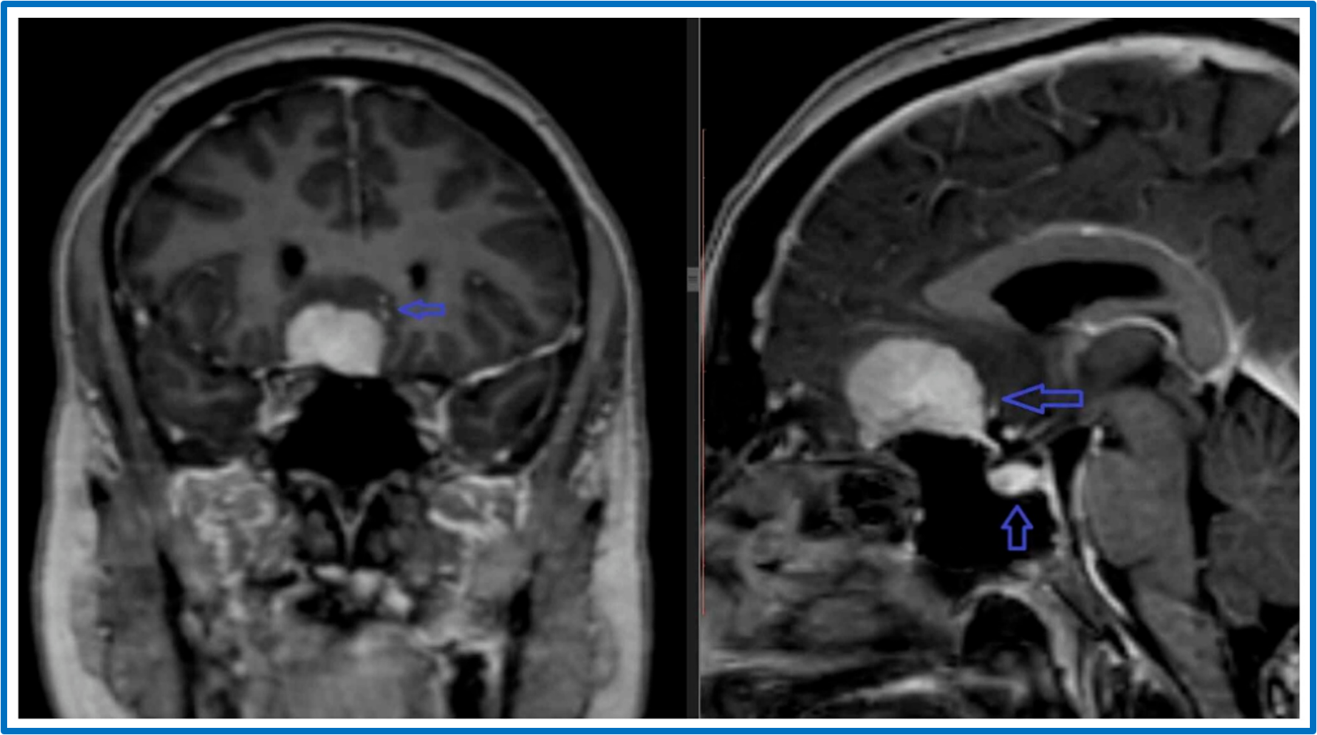
Suprasellar meningioma
Post-contrast MR images demonstrating coronal (left) and sagittal (right) views of an intensely enhancing extra-axial mass (top arrow) arising from the planum sphenoidale, with a dural tail visible on the sagittal view. The normal-appearing pituitary gland within the sella is also noted (bottom arrow).
Image credit: Adapted from Chaudhry S, Raza, Naveed MA, et al. Suprasellar Meningiomas: An Experience of Four Cases With Brief Review of Literature. Cureus. 2021;13(1):e12470. doi:10.7759/cureus.12470. Licensed under the Creative Commons Attribution 4.0 International (CC BY 4.0) licence. Modifications include [describe changes]. Title and caption modified.
Clinical reminder: consider NECSA mechanisms
(Neuropsychiatric Effects of CNS Structural Abnormalities)
When a structural lesion is identified in a patient presenting with neuropsychiatric symptoms, consider which NECSA mechanisms may explain these symptoms:
● Network disruption
Disruption of functional networks due to the presence of the lesion
● Electrical disturbance
Ictal, preictal, postictal, or interictal effects due to epileptic activity associated with the lesion (particularly if involving or adjacent to cortex)
● Chemical (endocrine) effects
Hormone excess or deficiency caused by the lesion (particularly if involving the pituitary gland or hypothalamus)
● Side effects of treatment
Effects of medications, surgery, radiotherapy, or other interventions
● Alternative explanations
The lesion may be incidental, or symptoms may arise from another cause (e.g., psychological response to illness or a comorbid psychiatric condition)
Selected references and further reading — Lesional lens
Rohlfing T, Zahr NM, Sullivan EV, Pfefferbaum A. The SRI24 multichannel atlas of normal adult human brain structure. Hum Brain Mapp. 2010;31(5):798–819.
Clark DL, Boutros NN, Mendez MF. The brain and behavior: an introduction to behavioral neuroanatomy. 4th ed. Cambridge: Cambridge University Press; 2018.
Brazis PW, Masdeu JC, Biller J. Localization in clinical neurology. 7th ed. Philadelphia: Wolters Kluwer/Lippincott Williams & Wilkins; 2016.
Blumenfeld H. Neuroanatomy through clinical cases. 3rd ed. New York: Oxford University Press; 2021.
Hassan I. The neuropsychiatric effects of CNS structural abnormalities (NECSA) classification: an aid to differential diagnosis. Aust N Z J Psychiatry. 2015;49(10):943.
Arciniegas DB, Yudofsky SC, Hales RE, editors. The American Psychiatric Association Publishing textbook of neuropsychiatry and clinical neurosciences. 6th ed. Washington (DC): American Psychiatric Association Publishing; 2018.
Agrawal N, Faruqui R, Bodani M, editors. Oxford textbook of neuropsychiatry. Oxford: Oxford University Press; 2020.