
Thalamus
Page most recently updated 7 September 2026
This page focuses on the neuropsychiatric manifestations associated with lesions of the thalamus. The thalamus is a major hub integrating widespread cortical and subcortical networks underlying sensory processing, arousal, cognition, memory, and emotion.
Cognitive, behavioural, and psychiatric effects of lesions may also arise through mechanisms other than network disruption; the potential contribution of these mechanisms can be considered using the Neuropsychiatric Effects of CNS Structural Abnormalities (NECSA) classification.
On this page:
Tuberothalamic artery (anterior) territory
Paramedian artery territory
Inferolateral artery territory
Posterior choroidal artery (posterior) territory
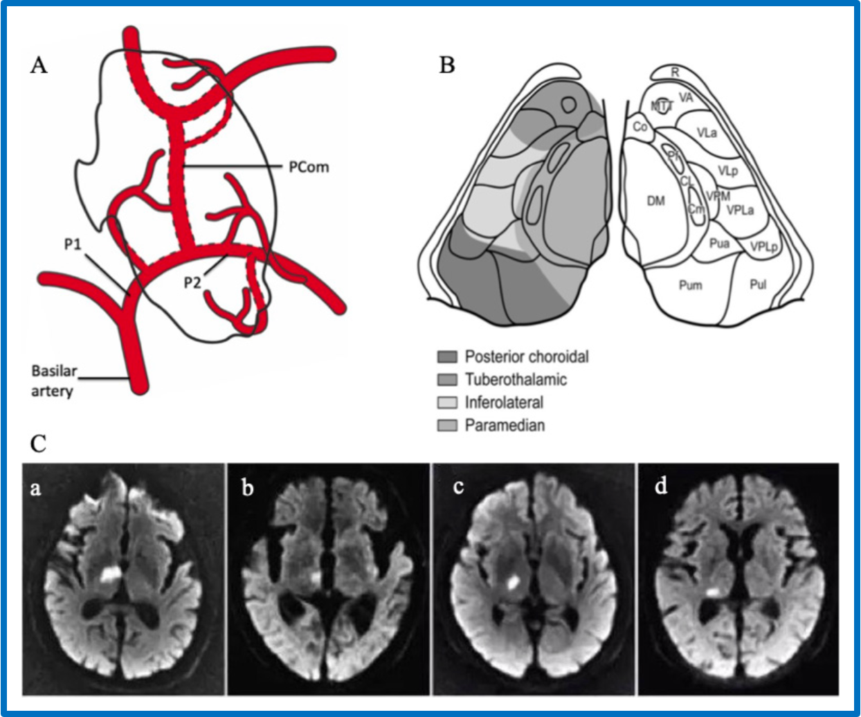
Thalamic arterial territories, nuclei, and infarction patterns
A. Illustration of the major arterial supply of the thalamus. B. Axial-plane illustration showing the thalamic nuclei and their corresponding arterial territories. C. Diffusion-weighted MRI images from four patients, each demonstrating an infarct in the (a) anterior territory, (b) paramedian territory, (c) inferolateral territory, and (d) posterior territory.
Image credit: Reproduced unchanged from Ye C, Pan R, Wu B. Improving the understanding of neural mechanisms and guiding targeted therapy for clinical syndrome after thalamic infarction with advanced neuroimaging. Human Brain. 2023;2(1). doi:10.37819/hb.1.307. Licensed under the Creative Commons Attribution-NoDerivatives 4.0 International (CC BY-ND 4.0) licence.
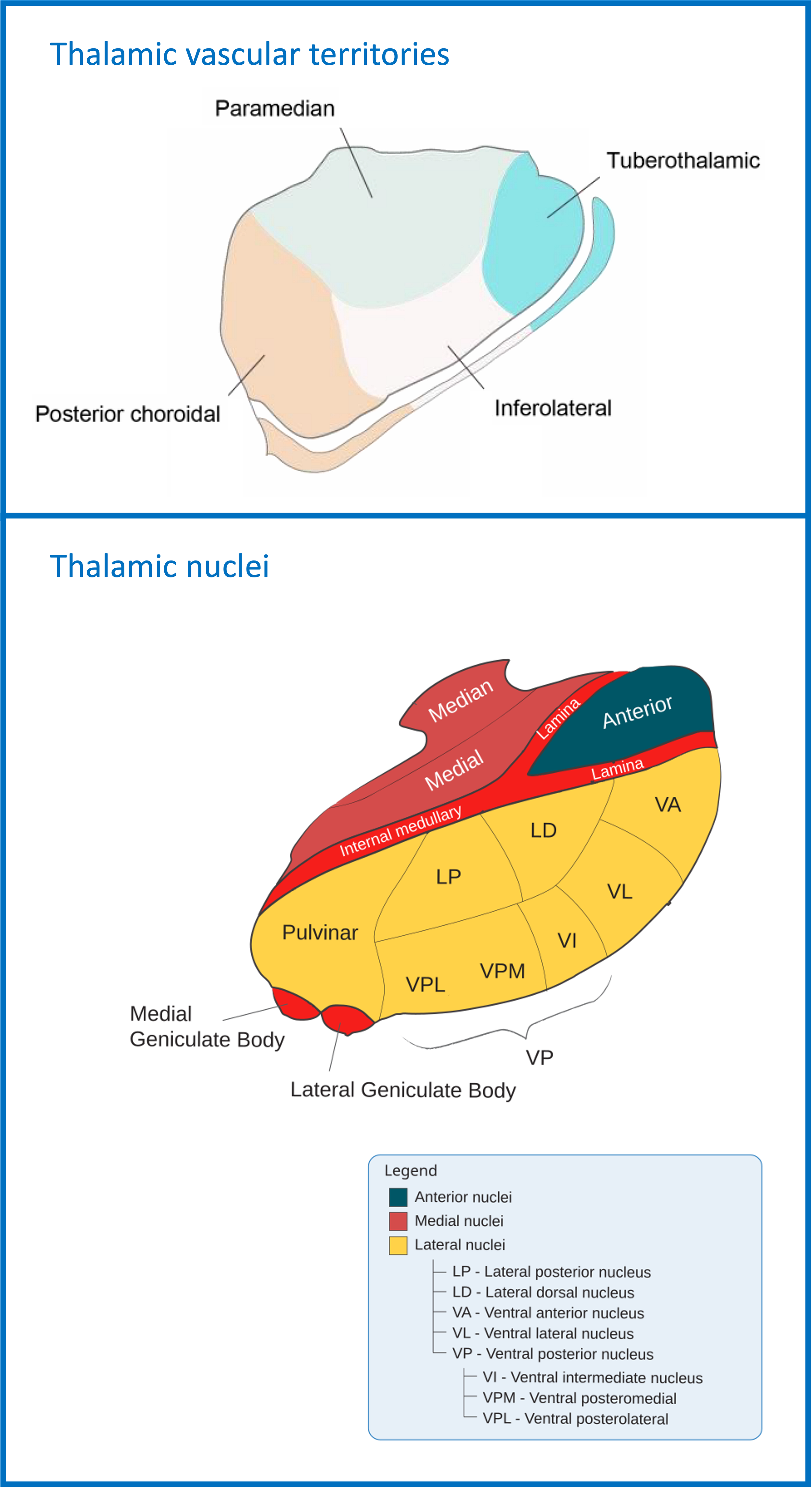
Thalamic arterial territories and nuclei
The upper diagram illustrates the arterial territories of the thalamus, while the lower diagram shows the nuclei of the thalamus. Note that the mediodorsal nucleus is not labelled in the diagram but occupies much of the territory labelled as medial.
Image credit: Upper image adapted from “Talamo territorios vasculares.png” by Irina Filchenko, Claudio L. A. Bassetti, and Carolina Gutierrez Herrera via Wikimedia Commons. Licensed under the Creative Commons Attribution 4.0 International (CC BY 4.0): https://creativecommons.org/licenses/by/4.0/. Lower image adapted from “Thalamus-schematic.svg” by Madhero88 and Jon C via Wikimedia Commons. Licensed under the Creative Commons Attribution–ShareAlike 3.0 Unported (CC BY-SA 3.0): https://creativecommons.org/licenses/by-sa/3.0/. Composite figure created by combining and modifying images from the above sources. Title and caption modified. The composite figure is licensed under the Creative Commons Attribution–ShareAlike 4.0 International (CC BY-SA 4.0).
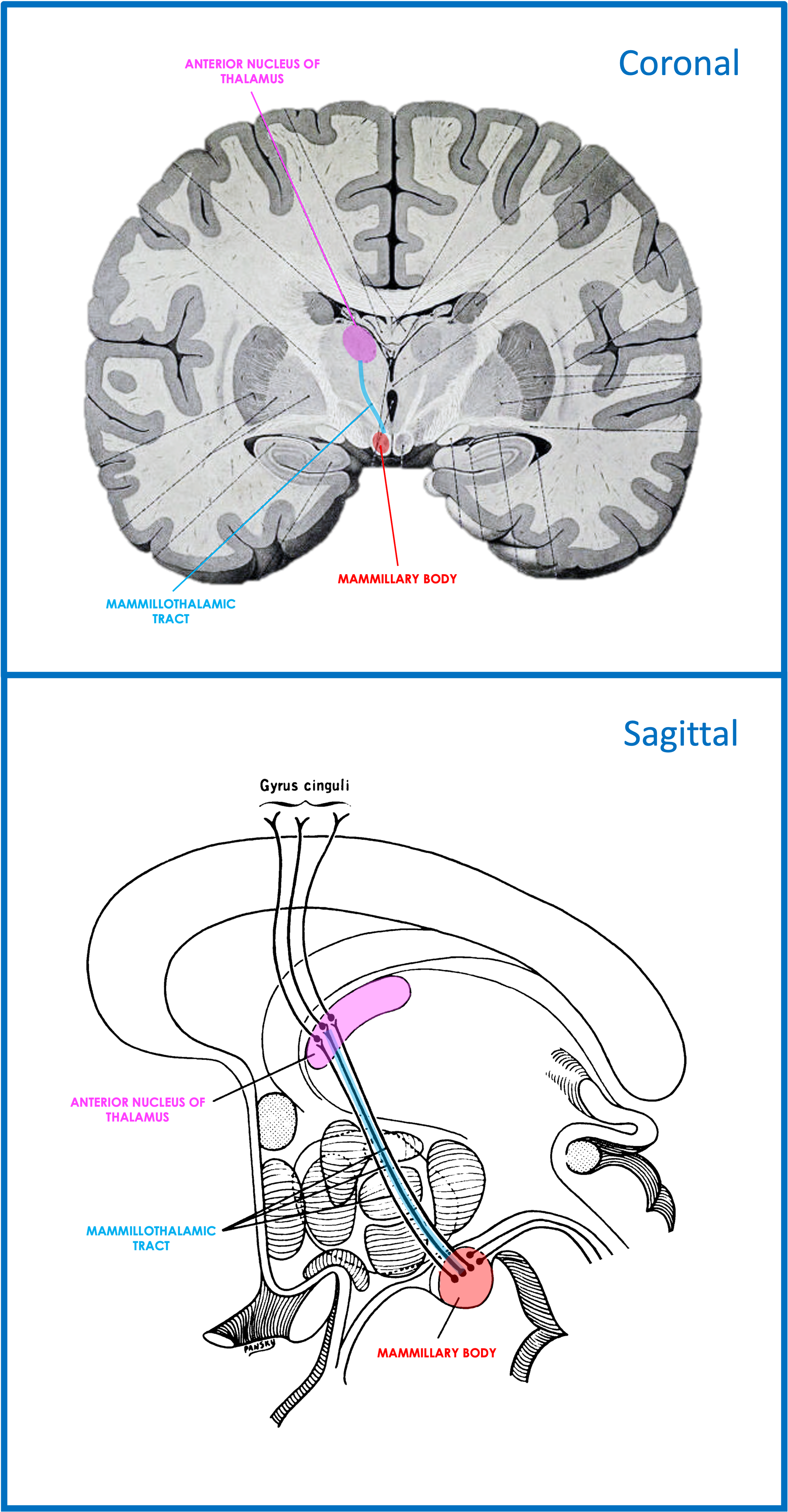
Mammillothalamic tract
The upper panel shows a coronal view of the mammillothalamic tract, extending from the mammillary body to the anterior nucleus of the thalamus. The lower panel provides a sagittal schematic view of the same pathway.
Image credit: Upper image adapted from Sobotta's Atlas of Human Anatomy, edited from the 8th German edition by J. Playfair McMurrich (1930), via the Wellcome Collection. Public Domain Mark. Lower image adapted from "Lawrence 1960 21.7.png" via Wikimedia Commons. Licensed under the Creative Commons Attribution–ShareAlike 4.0 International (CC BY-SA 4.0): https://creativecommons.org/licenses/by-sa/4.0/. Composite figure created by combining and substantially modifying images from the above sources. Title and caption modified. The composite figure is licensed under the Creative Commons Attribution–ShareAlike 4.0 International (CC BY-SA 4.0).
Thalamic vascular territories correspond to characteristic patterns of nuclei involvement and associated neuropsychiatric manifestations:
1. Tuberothalamic artery (anterior) territory:
Lesions involving the anterior nucleus (AN) may cause the anterior behavioural syndrome, characterized by reduced initiation of speech and behaviour (apathy), as well as anterograde amnesia and confabulation. The amnestic syndrome may relate to disruption of mammillothalamic connections. Left-sided lesions involving adjacent language-related thalamic nuclei may produce thalamic aphasia, while right-sided lesions may be associated with hemispatial neglect.
2. Paramedian artery territory:
Mediodorsal (MD) nucleus lesions may cause executive dysfunction and memory impairment. Right-sided MD nucleus lesions have also been associated with behavioural disinhibition that may mimic mania. MD nucleus involvement commonly occurs as part of broader paramedian thalamic lesions, which can additionally cause vertical gaze paresis, hypersomnolence, and diffuse central post-stroke pain syndromes. Decreased arousal and unresponsiveness may be marked in patients with bilateral paramedian infarcts. Hypersomnolence and central post-stroke pain syndromes may relate to involvement of the intralaminar nuclei.
3. Inferolateral artery territory:
Inferolateral thalamic lesions involve the ventral lateral (VL), ventral posterior lateral (VPL), and ventral posterior medial (VPM) nuclei. VL nucleus involvement may disrupt cerebellothalamic pathways and contribute to the cerebellar cognitive affective syndrome (hyperlink), as well as contralateral hemiataxia. VPL and VPM nucleus lesions may cause contralateral sensory loss and regional central post-stroke pain, particularly with limb involvement.
4. Posterior choroidal artery (posterior) territory:
This territory, including the pulvinar, is not emphasized in this section because it is primarily associated with visual attention and perceptual functions rather than prominent behavioural syndromes.
Clinical reminder: consider NECSA mechanisms
(Neuropsychiatric Effects of CNS Structural Abnormalities)
When a structural lesion is identified in a patient presenting with neuropsychiatric symptoms, consider which NECSA mechanisms may explain these symptoms:
● Network disruption
Disruption of functional networks due to the presence of the lesion
● Electrical disturbance
Ictal, preictal, postictal, or interictal effects due to epileptic activity associated with the lesion (particularly if involving or adjacent to cortex)
● Chemical (endocrine) effects
Hormone excess or deficiency caused by the lesion (particularly if involving the pituitary gland or hypothalamus)
● Side effects of treatment
Effects of medications, surgery, radiotherapy, or other interventions
● Alternative explanations
The lesion may be incidental, or symptoms may arise from another cause (e.g., psychological response to illness or a comorbid psychiatric condition)
Selected references and further reading — Lesional lens
Rohlfing T, Zahr NM, Sullivan EV, Pfefferbaum A. The SRI24 multichannel atlas of normal adult human brain structure. Hum Brain Mapp. 2010;31(5):798–819.
Clark DL, Boutros NN, Mendez MF. The brain and behavior: an introduction to behavioral neuroanatomy. 4th ed. Cambridge: Cambridge University Press; 2018.
Brazis PW, Masdeu JC, Biller J. Localization in clinical neurology. 7th ed. Philadelphia: Wolters Kluwer/Lippincott Williams & Wilkins; 2016.
Blumenfeld H. Neuroanatomy through clinical cases. 3rd ed. New York: Oxford University Press; 2021.
Hassan I. The neuropsychiatric effects of CNS structural abnormalities (NECSA) classification: an aid to differential diagnosis. Aust N Z J Psychiatry. 2015;49(10):943.
Arciniegas DB, Yudofsky SC, Hales RE, editors. The American Psychiatric Association Publishing textbook of neuropsychiatry and clinical neurosciences. 6th ed. Washington (DC): American Psychiatric Association Publishing; 2018.
Agrawal N, Faruqui R, Bodani M, editors. Oxford textbook of neuropsychiatry. Oxford: Oxford University Press; 2020.